Suspected PE 1 6 811. Up to 5 ccsec.
Ct Pulmonary Angiogram Wikipedia
Once the time to peak is determined ex.

Ct pe protocol. Protocol for 16-Section CT of Pulmonary Embolism Scanning delay is determined by dividing the acquisition time for lung imaging by 2 and subtracting the. Modern MDCT scanners are. Ad Full Comprehensive CT Lung Scan Based in Maryland.
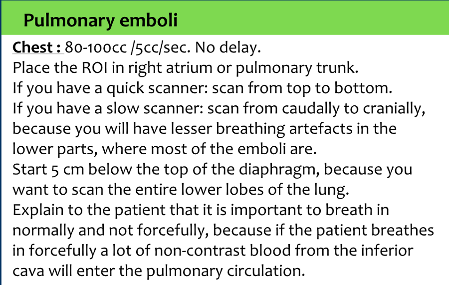
Rescan lower abdomen and pelvis. Adequate enhancement of the pulmonary trunk and its branches. For a pulmonary embolism CT an ROI is placed in the pulmonary artery and a test bolus is given.
100 cc of Isovue 300 contrast in a 1 liter saline bag The Foley catheter must be placed by the trauma or emergency service who should have already cleared the patient from possible urethral injury. There are two principal approaches for performing a. Entiate between true pulmonary embolism and a variety of patient-related technical anatomic and pathologic factors that can mimic pulmonary embolism.
6 seconds the scan is set to initiate the full scan 6 seconds after the injection of the main contrast bolus. Instruct the patient to take shallow breaths not deep breaths. Accurate interpretation of post-operative abdominal CT scans requires an understanding of the normal post-operative anatomy as well as potential complications of each.
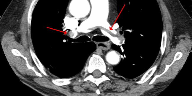
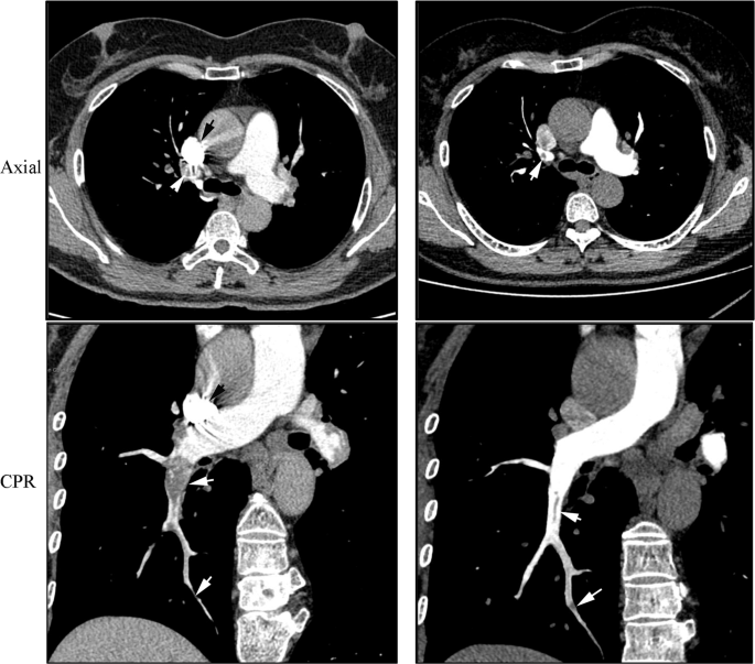
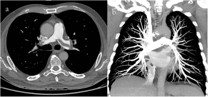
CT pulmonary angiogram is a medical diagnostic test that employs computed tomography angiography to obtain an image of the pulmonary arteries. Currently the most commonly used first-choice imaging examina-tion in patients with suspected PE is pulmonary CT angiography CTA 7. Or at 70 sec pi.
Ad Full Comprehensive CT Lung Scan Based in Maryland. Accurate and reliable diagnosis of acute pulmonary embolism PE is crucial to enable rapid treatment and guide patient management Pulmonary CT angiography CTA has been firmly established as the modality of choice in suspected acute PE 2 3Owing to technical advancements along with the immediate and widespread availability of this test pulmonary CTA has long superseded pulmonary. Pulmonary Embolism CT Perfusion.
This recommenda-tion is based on high sensitivity and specificity for PE and other clinically important conditions that mimic PE eg cardiac failure pneumonia pneumothorax. Protocol for 16-Section CT of Pulmonary Embolism Parameter Normal-sized Patients Large Patients 250 lb Detector width mm 125 25 Reconstruction interval mm 0625 125. Evaluate for hypovascular mets.
Nc art ven 5 min. Instruct patient not to contract or tense up their stomach muscle when they hold their breath. Pulmonary Embolism Standard Protocol.
Please make sure it states Power injectable. It is a preferred choice of imaging in the diagnosis of PE due to its minimally invasive nature for the patient whose only requirement for the scan is an intravenous line. Dilute contrast is a 2-3 solution of iodine.
RN MD PA. Its main use is to diagnose pulmonary embolism. Flow up to 5mlsec up to 300 PSI.
Right Coronary Artery Stent. Please make sure it states Power injectable. Each radiology department will have a slightly different method for achieving the same outcome ie.
Art ven 5 min. Stenosis of the Left Anterior Descending Artery. Test flush for ease of flushing and proper flow to achieve bolus injection no more than 5ccsec.
2 CT Protocols CT Protocols IV Contrast Indications Massmalignancystaging May require a special multiphase protocol InfectionInflammation Pain Unsure Angiograms Contraindications Allergy GFR30 45 Caution in hypertension diabetes renal transplant single kidney CRD. CT protocols ranging from a simple noncontrast CT at one end of the spectrum and CT perfusion as a complex protocol available only on highend scannersWith the growing diversity there is. HCC 1st time screening or known hyperdense nodules.
It is a matter of personal flavor to do the whole abdomen at 35 sec pi. Pe protocol yes ct chest with iv contrast3d reconstruction for pe protocol can be coded as 71275 thanks kavitha s cpc. CT examination of the pancreas should always be done with maximum amount of contrast at a maximum flow rate because both small pancreatic carcinomas aswell as pancreatic necrosis in pancreatitis are difficult to detect.
Follow up or screening in HCC patient. When the patient takes the final breath in to hold instruct patients to take only a small breath in and then stopsuspend breathing. CT Tech and b.
Please see notes on page 3 in regards to Bard Provena Picc Lines. The computed tomography pulmonary angiogram CTPA CTPE is a commonly performed diagnostic examination to exclude pulmonary emboli.
 Ct Chest With Contrast Pe Protocol Ct Chest Pe Protocol Ct Showed A Large Thrombus Completely Occluding The Right Radiologic Technology Radiology Nerdy Nurse
 The Radiology Assistant Ct Contrast Injection And Protocols

 Ct Chest With Iv Contrast Pe Protocol Mild To Moderate Cardiomegaly Download Scientific Diagram
 Overuse Of Ct Scans For Suspected Pulmonary Embolism Persists Tctmd Com
 An Optimized Test Bolus For Computed Tomography Pulmonary Angiography And Its Application At 80 Kv With 10 Ml Contrast Agent Scientific Reports
Computed Tomography Ct Scan Of The Chest With Contrast Ct Scan Of Download Scientific Diagram
 Ct Imaging For Suspected Pulmonary Embolism Pt 1 Youtube
 Ct Angiography Of Pulmonary Embolism Diagnostic Criteria And Causes Of Misdiagnosis Radiographics
 Ct Pe Protocol Pneumothorax Page 1 Line 17qq Com
 Ct Scan Of The Chest With Pe Protocol Download Scientific Diagram
 Inventive Protocols Of Ct Pulmonary Angiography Ctpa Avoid Artifacts In Right Pulmonary Artery Rpa Improving Detectability Of Pulmonary Embolism Pe European Respiratory Society
 Use Of Pulmonary Ct Angiography With Low Tube Voltage And Low Iodine Concentration Contrast Agent To Diagnose Pulmonary Embolism Scientific Reports

No comments:
Post a Comment
Note: Only a member of this blog may post a comment.