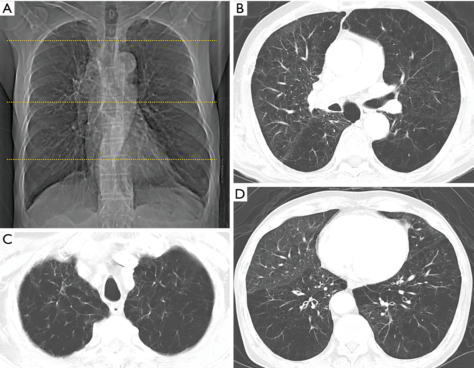
Remember that the left and right side are the opposite in a CT scan The reason for the extreme pneumothoraxlung collapse is not known. Doctors can see these dilated blood vessels on CT scan images.
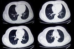
 Copd Restrictive Lung Disease Perokok O
Also the diaphragm may.
What does copd look like on a ct scan. This condition known as vascular dilation usually appears in the area of ground glass opacities. Arrows show dark areas in periphery of the lung with no blood vessels. Collapsed right lung and treatment resul.
Cancerous nodules grow extremely quickly. 11 With the introduction of clinical CT. Of the patients with false positives on CT nearly 7 had a more invasive diagnostic test such as a biopsy or bronchoscopy in which a scope is used to look down the airway to see if there is a mass.
There is no narrowing or obstruction of your breathing tubes airways. This means the lungs appear larger than normal. The following picture shows a collapsed right lung which is visible as a bright white area where the right lung should be.
Another lung disease that falls under the scope. Noncancerous nodules can look just like the cancerous ones However noncancerous nodules tend to be small and they grow very slowly if at all. If a CT scan is not available the presence of emphysema can be suggested by a low value for a breathing test called diffusing capacity.
COPD is quite often diagnosed just via spirometry but CT is the gold standard. I asked last time if you had obstruction on your spirometry and eventually assumed you didnt. Heres a look at signs you may have the emphysema or COPD.
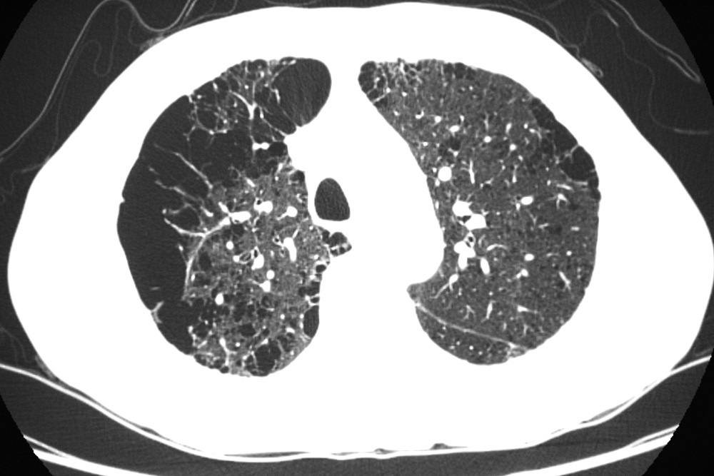
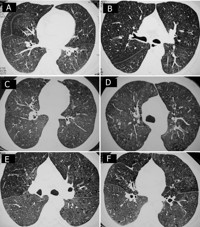
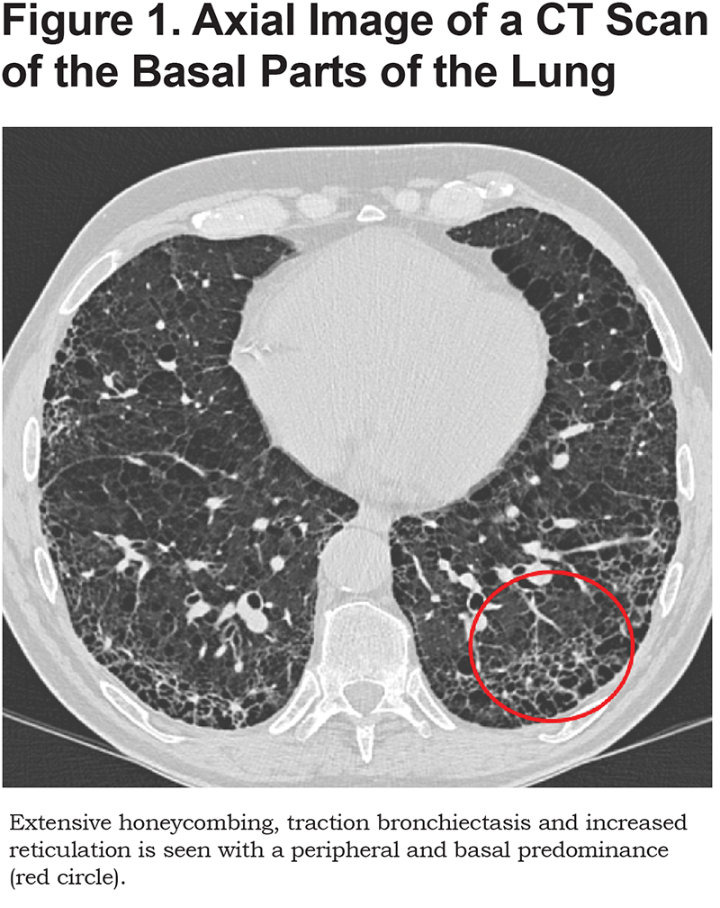
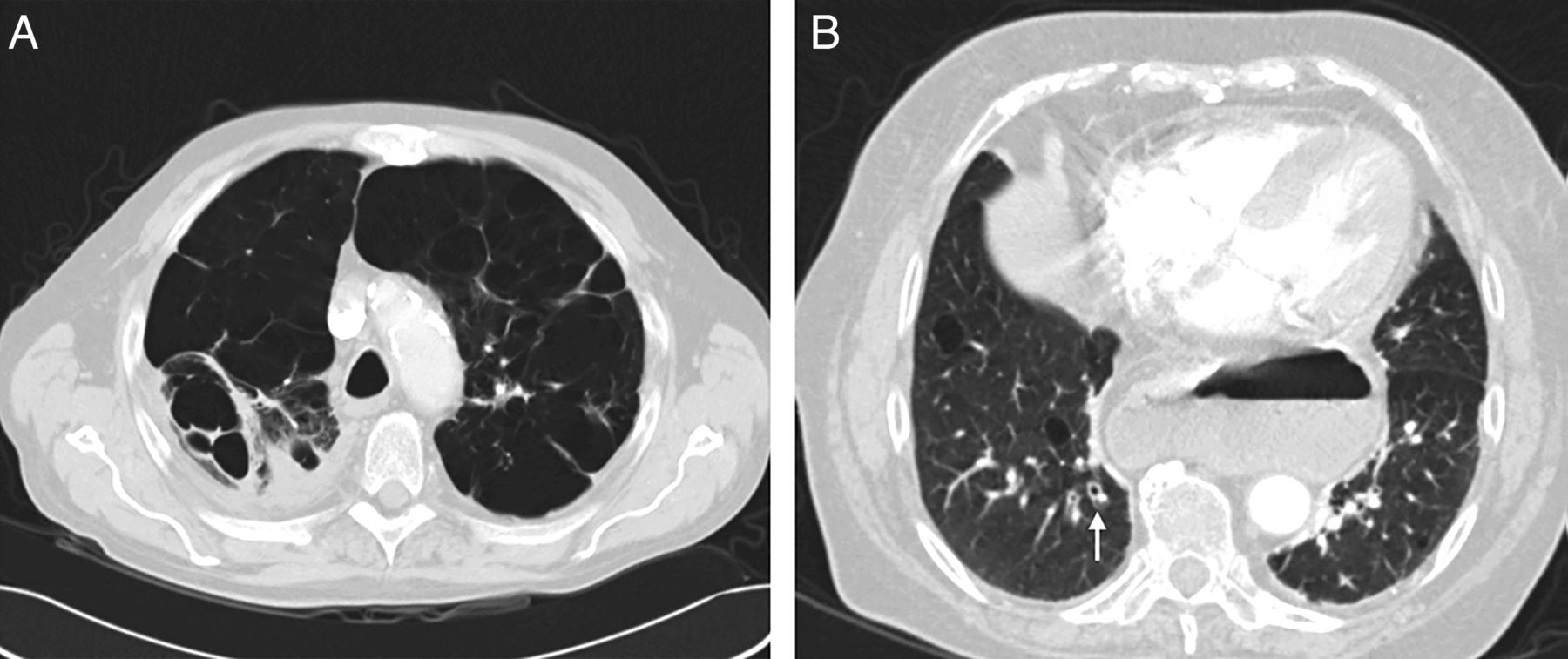
Additionally a high resolution CT scan is also excellent at detecting and determining the severity of bronchiectasis. A typical CT scan showing this type of COPD is shown below. How Doctors Identify Emphysema.
Diagnosing chronic obstructive pulmonary disease can involve an X-ray which may show enlarged lungs and diaphragm problems. Upper lung lower lung diffuse 10 small airway disease airtrapping bronchial wall dilatation wall thickening as well as large airway disease. Vascular dilation appears to play a role in hypoxemia in patients with COVID-19-related pneumonia.
A CT scan can pick up characteristics that a normal X-ray can miss like specific damage to the lungs directly caused by emphysema small lung nodules or even small lung cancers. CT scan shows emphysema in the left lung. The pneumonia typically appears along the walls of each lobe of the lung especially the chest wall and the lower portions of the lungs.
The CT picks them up as tissue that is different than the surrounding area which is why they light-up white on the images and are easier to see. A chest CT scan is a particular test used to diagnose COPD usually following an initial chest X-ray. Microscopic view of the air sacs alveoli in the top right showing emphysema destruction and enlargement.
Chest tightness or chest pain. Your doctor needs to do repeated CT scans over a long period to monitor the growth rate. Also since you are 35 yo you are at l.
Without obstruction you wont get a CT scan looking for COPD because you cant have COPD without obstruction. They can double in size every four months so your doctor will conduct repeated CT scans. One of the signs of COPD that may show up on an X-ray are hyperinflated lungs.
A cough that wont go away. During a CT scan of the chest detailed pictures are taken of cross sections or slices of the thoracic structures in your body. Thoracic structures include your lungs heart and the bones around.
Even the slightest activities may tire you out. If you have compromised lung function COPD that would put you at higher risk not some scarring seen on CT. A CT or CAT scan is a shortened name for computerized tomography.
CT scan of the chest. Centrilobular panlobular paraseptal 9 10 the presence of bullae 10 and their location ie. Many patients may have the disease but be unaware of it.
Some of the white spots are ACC nodulesmets. The earliest symptom of emphysema is shortness of breath. The video scrolls through the image slices of the scan and shows the typical white ground glass opacities GGO caused by COVID pneumonia.
X-rays can show irregularities or damage in the lungs caused by COPD and other acute and chronic lung diseases. They appear as round white shadows on a chest X-ray or computerized tomography CT scan. Using Xray CT the characterization of COPD has evolved beyond the traditional distinctions of emphysema and chronic bronchitis based on the presence and type of emphysema ie.
CT scan shows emphysema in the left lung. Coronavirus infection can widen or dilate the blood vessels of the lungs. This painless imaging exam is more detailed than a chest X-ray taking numerous cross-sectional pictures of the lungs and other organs inside the chest as well as blood vessels and bones from various angles which are then combined by a computer into 3-D images.
Remember that emphysema is one of two types of COPD the other type is chronic bronchitis coughing up mucus most days. What does emphysema look like.
High Resolution Computed Tomography And Chronic Obstructive Pulmonary Disease Intechopen
![]() Chest Ct Scan Showing Emphysema And Nodule Arrow Download Scientific Diagram
Chest Ct Scan Showing Emphysema And Nodule Arrow Download Scientific Diagram
 Emphysema On Ct Scan Of Chest Youtube
 Incidental Findings On Chest Ct Imaging Are Associated With Increased Copd Exacerbations And Mortality Thorax
 Quantitative Ct Assessment Of Chronic Obstructive Pulmonary Disease Radiographics
 Thin Section Ct Scan Of A Smoker With Chronic Cough Associated With Download Scientific Diagram
 Advanced Imaging In Copd Insights Into Pulmonary Pathophysiology Milne Journal Of Thoracic Disease
 How Are Ct Scans Used In Detecting Copd Copd Foundation

 Chronic Obstructive Pulmonary Disease Radiology Reference Article Radiopaedia Org
 Prognostic Significance Of Ct Determined Emphysema In Patients With Small Cell Lung Cancer Lee Journal Of Thoracic Disease
 Copd Bronchiectasis Overlap Syndrome European Respiratory Society
 Ct Definable Subtypes Of Chronic Obstructive Pulmonary Disease A Statement Of The Fleischner Society Radiology


No comments:
Post a Comment
Note: Only a member of this blog may post a comment.